Showing posts with label NCLEX cramming. Show all posts

Tuesday, January 19, 2016

Day 7: Health Screening and Disease Prevention (Contraception, STIs, and Antibiotics), & Eating Disorders, Violence, Abuse, and Assault

HEALTH SCREENING & DISEASE PREVENTION

There are 3 levels of health prevention:

  • primary: preventing the disease entirely (immunizations, nutrition and fitness, dental cleaning, safety features eg. seatbelt)
  • secondary: focusing on identifying the disease, treating it, and preventing the spread (condoms, isolation, etc.)
  • tertiary: focus on treating the long-term consequences of chronic illnesses/disability for optimal quality of life (rehabilitation, prevention of ulcers, medication, etc.)


Screening guidelines for everyone:

  • dental q 6 mos
  • BP @ 20 y.o. should be 120/80, screen yearly, biannual if higher than 120/80.
  • BMI annual.  Remember, BMI= kilos/(m^2) or (730 x lbs)/(in^2)
  • Cholesterol<200 q 5 years starting @ 20 y.o.
  • Glucose 90-120, A1C (4-5) q 3 years starting @ 45 y.o.
  • Colorectal:  

annual fecal occult beginning @ 50

& sigmoidoscopy q 5 years

or colonoscopy q 10 years

or double contrast barium q 5 years
or CT colonography q 5 years


For women: 
  • breast exam q 3 years starting @ 20, then annual starting @ 40 y.o.
  • pap q 3 years starting @ 21
  • annual mammogram starting @ 40
There are some female reproductive diseases that do not have good options for screening, such as 
  • Ovarian cancer (risks include: first pregnancy after 30 y-o, family hx of ovarian, breast, colon cancer, hx of dysmenorrhea, use of hormone therapy/infertility medications, and age over 40 y-o.), which has vague symptoms like pain and pressure of the abdomen, pelvis, back, or legs, GI symptoms, fatigue, frequent urination, or vaginal bleeding.  Treatment is surgery, chemo, radiation.
  • Myomas (uterine fibroids) have very similar sign and symptoms of pelvic pain and pressure and hypermenorrhea.  Treatment is surgery.
  • Endometriosis also have lower abdominal pain and dysmenorrhea.  It causes pain during sex, and back and rectal pain.  Treatment is oral contraceptives or surgery.
  • Endometrial cancer causes vaginal bleeding between menstrual cycles or after menopause, and the risk increases three-fold for women who are 21+ pounds overweight.  Treatment is hysterectomy, radium, and x-ray therapy.

For men:
The Try Guys Get Prostate Exams


CONTRACEPTIVES

So the basic principle of birth control is either tricking your body into thinking its pregnant (which prevents follicular development), or preventing the sperm from meeting the ova.  Everything else is just variation.  You have abstinence, sterilization, or rhythm method, or coitus interruptus, which all attempt to do the same thing, which is to prevent the sperm from meeting the egg.  
Then you have hormonal treatments, whether in depot form (depot medroxyprogesterone acetate aka DepoVera, patch, pill, ring, or even IUD.  Then you have spermicidal substances in various methods of delivery and blocking the path of sperm, such as cervical capping, diaphragm, condoms, and just pushing that spermicide into the vagina.


There's a lot of non-barrier, non-medication methods for birth control, but I'm only going to talk about the rhythm method (aka Billings or Creighton method) which is pretty cool, but does require persistence (women's cycle do fluctuate with just about any type of stress-illness, work, etc.) Basically the woman monitors cervical mucus changes when toileting (watery mucus=ovulation because sperm needs to be able to get through the cervical os).  Also you record your menstruation, and basal temperature.  A temperature spike means LH surge, which means ovulation and then you have 12-24 hours to get jiggy with it to make a baby.  (Well, technically sperm is viable around 72 hours to 5 days, so you have a bigger window in terms of having sperm floating around for a while before you ovulate.)  Btw, there's a plethora of fertility apps to help you track your cycle for whatever reason.  I'm a fan of Kindara.  


Hormone method

Progesterone and estrogen contraceptives:
Basically estrogen and progesterone hormones causes the hypothalamus and anterior pituitary to shut down from negative feedback.  Contraindications for this include a history of thrombosis or being in a high risk for thrombosis at this time, estrogen receptor positive tumors, lactation that is less than 6 weeks postpartum, and liver tumor.

Progestin-only contraceptives:
Causes inhibition of ovulation, thickening the cervical mucus, thinning the endometrium, and altering the cilia in uterine tubes.  The main thing to remember about progestin-only methods is that because they don't contain estrogen (estrogen suppresses lactation), they can be used during lactation.  You also have to remember that it has to be taken at the same time every day, and directions for making up a missed dose will vary.  If you miss 2 or more in a row, you should get back on but also use the Morning After pill.

Method of delivery
Oral contraceptives:  Can come in progestin only or estradiol/progestin remember to take it at the same time every day, and that if you miss one pill to just take it as soon as possible, and if you miss 2 days, use a backup method for a week.  Hormonal contraceptives decrease the efficacy of anticonvulsants, antifungals, antituberculosis, and anti HIV medications -- or rather, I should say, any medications with the suffix anti- in it will decrease the efficacy of contraceptives.  SE: breast tenderness, bleeding, n/v.  Avoid smoking.

Ethinyl estradiol and norelgestromin/transdermal patch: comes in the estradiol/progestin combination and you wear it on the low abdomen, upper outer arm, buttock or chest (not breasts).  Replace on the same day every week for 3 weeks, rotating sites, with one week without a patch for menstruation.

Levonorgestrel 1.5 mg or "Plan B":
 aka the morning after pill is made of estradiol/progestin combination and comes in a larger than normal dose of oral contraceptive that has to be taken within 72 hours of unprotected sex.  The next pill should be taken 12 hours after the first pill.  This should never be taken as a primary method of birth control.
  
NuvaRing: comes in estradiol/progestin and is placed deep into the vagina (most comfortably around the cervix, but it's not necessary for perfect placement) for 3 weeks, then removed to allow for menstruation during the 4th week.  This doesn't have to be fitted, but some wearers may experience vaginal discomfort/increased vaginal discharge.  Sometimes when a woman bears down (bowl movement or whatnot) it may shift downward and protrude from the vagina.  All you need to do is wash it and put it back up around the cervix within 5 hours of it coming out.  Though it's not recommended to remove it for intercourse, the effectiveness will not decrease if replaced within 3 hours.  It has the same rate of efficacy as the other routine birth controls (pill, condoms, etc.).

Intrauterine device:  Here's a video about IUDs.  They have to be placed and removed by a doctor, and they are 99% effective if correctly placed.  There are two types of IUDs: hormonal or copper.  Both types cause inflammation of the uterine lining which triggers immune cell release that will kill sperm, but the hormone IUD also causes thickening of mucus in the cervix and thins the endometrial lining so it is less likely to receive a blastocyst.  The hormone reservoir lasts 7 years.  The copper IUD lasts 12 years.  The string of the IUD is just there to reassure a woman that it is still in place.  Since it causes inflammation, it's a bad idea to use this if you have a pelvic infection.  You need to teach women to check for the IUD thread after their period to make sure that the IUD hasn't be expelled with the menstruation debris.  Also, check for copper allergies.  If a pregnancy does occur with the IUD in place the IUD needs to be removed immediately, and an U/S needs to be done to rule out placenta previa or ectopic pregnancy.  Also check for septic shock as there is a high risk of infection with IUDs.

Progestin only depot aka Depo-Provera:  given SQ or IM.  It is given during the first 5 days of the menstrual cycle and then administered every 11-13 weeks.  You have to use a backup form of birth control for 7 days after the first injection.  Return to fertility occurs about a year after cessation, but may be delayed as long as 18 months after discontinuation.  Side effects include osteoporosis, weight gain, increased thrombosis, irregular spotting, decreased libido, and breast changes.


Spermicidal Method with Mechanical Blocking

Method of delivery
Cervical cap: the "FemCap" is the only cervical cap available in the U.S. You can watch an instructional video here.  It's made of silicone and comes in three sizes.  You insert it with the taller brim dorsally, and the shorter brim anteriorly.  You put spermicide on the inside bowl that will be put over the cervix, around the brim, and on the dome.  Insert it with the bowl over your cervix (usually easiest in a sitting position like you would putting in a tampon--I would recommend a chair, though, not the toilet), and hold it for 10 seconds in place to create a seal 15 minutes before sexual arousal, and keep it there for at least 6 hours and no more than 48 hours at a time.  To remove, dimple in the dome to break the seal and pull it out by the strap.  Wash with water and soap, dry, and put it in the storage container.  You should get a pap smear every 3 months with the cervical cap.  You shouldn't use a FemCap if you are on your period, had an abortion within the last 6 weeks, gave birth less than 8-10 weeks ago, or if you have a uterine/cervix/vaginal infection or disease or an abnormal Pap smear.  And of course, you have to have familiarity, and it does take time, effort, and skill to insert.

Cervical diaphragm: basically work the same way the FemCap does. It can be made of rubber or silicone, and basically, you put spermicide inside of the dome, insert, and cap the cervix.  You need to have annual gynecological exams to assess the fit, especially after weight loss or gain of 10 lbs or more, births, or second trimester miscarriage, or after any abdominal or pelvic surgery.  It can be inserted 6 hours before sex and removed 6-8 hours after sex.  The main complaints is that it's difficult to insert, and that the cold diaphragm and cold gel can temporarily reduce the vaginal response if inserted immediately before intercourse.   It isn't a good option for women with poor vaginal muscle tone or recurrent UTIs.

Condom:  Check the expiration date, open (not with your teeth or you might tear the condom).  Makes sure you're putting the right side of the condom on your penis.  (Hint: a condom has a pointy bit, like a sombrero.  Put the sombrero on your penis like you would wear a hat.)

http://www.womenshealthmag.com/sites/womenshealthmag.com/files/images/bc-center-condoms-art.jpg
Pinch the pointy bit to make sure there's space for the sperm and use your other hand to roll the rest down the shaft.  When you've ejaculated, hold onto the edges of the condom as you withdraw from your partner while you are still erect, and then throw it away, don't just lay there until you get another erection and keep going.  You have to switch condoms between each ejaculation.  DON'T USE PETROLEUM BASED LUBRICANTS as this will break down the condom and decrease efficacy.  In fact you should only be using water based lubricants.  Do not use KY Jelly, it's made with glycerin, which increases risk of yeast infection--and trust me, you don't want a yeast infection.  The best part about condoms is that they prevent STIs when used properly.  Except not natural skin condoms--like lamb cecum--because the pores are big enough for viruses to pass through.

Here's a great website that was very practical about gay sex, but also just how to put on a condom. 

Female condom: basically, keep the rim on the outside of the vagina, and stick the part that looks like a collapsible cylinder into your vagina.  Boom.  Done.  And, I was talking to a friend who is a sex educator and podcaster about how female condoms are actually quite comfortable.  Here are articles about the product history, and how female condoms are the next big thing for safe sex.

Spermicide: nonoxynol-9 (N-9) reduces mobility of sperm upon contact.  It should be used no more than 1 hour before sexual intercourse, and inserted (a preloaded vaginal suppository, foam, or what have you) high up into the vaginal so that it contacts the cervix.  It also has to be reapplied for every additional romp (ok, ok, "act of intercourse").  Using it with a diaphragm or cervical cap increases efficacy.  Interestingly, results from studies show that the use of N-9 may increase transmission of HIV and cause lesions.  And that there is no evidence that the addition of spermicides to male condoms decreases the risk of pregnancy, so it is suggested that women with high HIV or STI risk behaviors avoid the use of spermicidal products containing N-9, which include the lubricated condoms.  (This was taken from the fifth edition of Perry, Hockenberry, Lowdermilk, and Wilson's "Maternal Child Nursing Care" page 125).  However, for the purposes of preventing pregnancy, you should definitely use a barrier method with N-9.

Mic's "How does birth control work?  Nobody seems to know."
Educator: Have you heard about IUDs?  
Man: Maybe you can fish with it?  For eggs?
^^ The reason why we need to have medically accurate sex education in schools.  


SEXUALLY TRANSMITTED DISEASES

GCCT=gonorrhea and chlamydia both mandatory report
  • gonorrhea short incubation of 3-8 days: clear or cloudy discharge, treated with ceftriaxone IM, and erythromycin ophthalmically with neonates (blindness).  
  • chlamydia aka "the silent disease" because s/s are absent or minor and the incubation is up to 1-3 weeks: yellow/green discharge.  (The ATI book also says "gray vaginal discharge."  Tell me, what's the difference between cloudy and gray?  Nobody knows?  Ok, then.)  Treated with azithromycin, doxycycline, and erythromycin ophthalmically with neonates (blindness)

Both cause:
  • vaginal bleeding, swollen testicles
  • frequent and painful urination/dysuria
  • diffuse/nonspecific abdominal pain
  • causes infertility and sterility
  • both cause complications of pelvic inflammatory disease

Both can be passed to neonate during childbirth, but gonorrhea can be passed in utero.  Often results in PID.

Pelvic inflammatory disease: Only occurs in women.  S/s chronic abdominal pain, green or yellow vaginal discharge with unpleasant odor and dysuria. Often accompanied by fever, chills, N/V.  Can lead to ectopic pregnancy and lead to infertility.  

Syphilis mandatory reportcan be passed in utero.  May cause miscarriage, heart disease, blindness, brain damage in neonate.  Treat with penicillin.  
Syphilis has 3 stages:
stage 1=sores (appears within 3 months of infection) lasting up to 6 weeks.
stage 2=rash on body & flu symptoms.
stage 3=neurological/cardiovascular complications

Trichomoniasis: in 3 days to 2 weeks after infection you will see COPIOUS FROTHY, MUCOPURULENT, MALODOROUS green yellow vaginal discharge, or white, watery penile discharge.  All of this accompanies your run of the mill dysuria and irritation.  Treatment is metronidazole.

Herpes: can be transmitted even with no visible lesions.  C/S indicated if active lesions present the 2 weeks before delivery, but also can be transmitted in utero.  Acyclovir is started 36 weeks gestation to prevent outbreak before delivery.
Painful blisters for up to 3 weeks with possible systemic aches, enlarged lymph nodes and fever.  Recurrences are preceded with shooting/tingling pain for hours to days before blister eruption.
No cure, but the symptoms can be shortened by use of any of the -cyclovirs.  

Human papillomavirus: Many strains.  Some cause condylomas that look flesh-colored of gray swelling, or cluster together like cauliflower, others cause cervical cancer.  May cause recurrent respiratory papillomatosis.  Treatment is imiquimod and podophyllin, and surgery to remove large warts.  The best treatment is HPV vaccines as early as 9 years old, but definitely recommended for 11-12 year old girls.

HIV: can lie dormant for years without symptoms.  Acute retroviral syndrome are non-specific with fever, malaise, lymphadenopathy, skin rash, but usually occur within the first few weeks before antibody tests are even positive. Can be spread during childbirth or during breastfeeding.  Often first spotted because of signs of immunosuppression due to depletion of CD4 lymphocytes (candidiasis, weight loss, diarrhea, flu-like malaise).  Treatment of obstetrical clients include zidovudine, nevirapine, and C/S 2 38 weeks.

Just so you know (since this is always a test question regarding HIPAA vs mandatory reporting to the CDC) these are the STIs you typically have to report to the CDC: 
  • Chancroid
  • Chlamydia trachomatis
  • Gonorrhea
  • Hepatitis B
  • Hepatitis C
  • Syphilis, including congenital syphilis

Infections that are concerning during Pregnancy and L&D:

GBS: NORMAL FLORA in vagina--NOT an STI.  Culture @ 35-36 wk.  Tx: IVPB PCN q 4 hr during labor.
Listeria (deli meats/hot dogs/soft cheeses/unpasturized/refrigerated or smoked seafood): bacteria found in soil, water, plants NOT an STI.  Causes premature labor, miscarriage, stillbirth

TORCH: 
Toxoplasmosis: NOT AN STI passed through cat feces and other animals, or garden soils.  Causes blindness, brain damage.
Other: (syph, varicella, parvovarius)
Rubella: causes cardiac, eye and ear disability, intellectual disability, liver and spleen damage.  Remember MMR vaccine for mom after birth, no pregnancy for 1 month after.  Children get the MMR @ 1 year and 4 years.
Cytomegalovirus (no treatment or vaccine): spread through saliva--may cause permanent hearing loss, vision loss, mental disability, microcephaly, lack of coordination, seizures, death.
Herpes infections: tx: acyclovir @36 wks, C/s if lesions visible within 2 weeks of delivery


INFECTION CONTROL

Things to remember:

Avoid placing clients on contact precautions with patients who are immunocompromised, with open wounds, or have anticipated prolonged lengths of stay.

Standard precautions (HepB, HepC, HIV<--only spread through blood and sexual contact) standard precautions can also include gown, mask, and goggles should the need arise--in other words, standard precautions can be upgraded to contact precautions if risk of blood and body fluids is high.  Like, you can wear a gown for someone who just has food poisoning.  The thing about standard precautions is that you have to treat all blood and body fluids as if they were contaminated.
Contact precautions (MRSA, C. diff, Hep A&E, Herpes, Carbapenem-resistant enterobacteriaceae, rotavirus, salmonella, shigellosis, staphylococcus, VRE<--these are all diseases that have diarrhea): isolated preferred but may cohort with the same organism.   Sterile gown, gloves and mask (when risk of splashing, spraying, coughing, spitting, etc. occurs).   Client leaves room only for essential clinical reasons.
Droplet precautions (meningococcal, pneumonia, RSV, rubella<--these diseases all have respiratory secretions that can be spread through coughing or sneeze): isolation preferred but may cohort with the same organism.  Door is kept closed.)  Must wear a mask within 3 feet of the patient.
Airborne precautions (varicella zoster/herpes zoster, rubeola, tuberculosis):  Requires individual negative pressure rooms.

Note: if these lists seem overwhelming to you, it's easier to simply memorize the droplet and airborne (there are fewer).

Surgical Asepsis: only dry items touch the field with 1 inch borders


EATING DISORDERS

Eating disorders are actually psychiatric disorders.  With eating disorders, the patient and the family may feel intense shame and are sensitive to others' perceptions.  You have to empathize with the patient's low self-esteem, unworthiness, and dysphoria, but be firm if the patient is dishonest, manipulative, or attempting to provoke you.  Reacting in an accepting and nonjudgemental way, and helping the patient to construct alternative ways to view their own behavior and logic will help to build trust.

There are 3 main types of eating disorders: anorexia nervosa, bulimia nervosa, and binge eating disorders.

Anorexia nervosa:
The thing to remember about anorexia is that the word "anorexia" is misleading because in medicine, the technical definition means "loss of appetite" as one might with chemo, or when nearing death.  A person with anorexia usually does have a n appetite.  Most anorexia patients restrict caloric intake, but some patients with anorexia actually do binge and purge--just not in cycles.  These patients often exhibit:

  • obsession with food (talking about it all the time whether about how good it tastes or how they can't eat something, but want to eat something, about the terror of gaining weight, etc.)
  • focused on food preparation and food handling (often pushing things around their plate to make it look like they're eating and eating for as long as everyone else at the table)
  • extreme exercise
  • self-induced vomiting, abuse of laxatives and diuretics (remember, electrolyte imbalance, especially hypokalemia)
  • cognition and self-perception impairment/judging self-worth by weight
immediate nursing concerns and s/s are


  • acknowledge the emotional and physical difficulty the patient is experiencing
  • hypokalemia, hypocalcemia, hypophosphatemia, hyponatremia--hypo everythingemia (CBC including hypoalbuminemia leading to edema)-->acute renal failure from dehydration--> cardiac dysrhythmias
  • muscle weakening, constipation
  • decreased fat, decreased estrogen, decreased calcium intake--> low bone density, amenorrhea
  • cold extremities-->lanugo

later nursing tasks are:

  • daily weights in bra and panties only with NO ORAL INTAKE before weighing--do not negotiate the resulting weight or reweigh
  • normalizing eating patterns
  • allow the patient to express feelings, and be encouraging, but remain firm about eating a set amount
  • preventing the patient from purging for 1 hour after eating, administer liquid supplements.
  • monitor bathroom trips after visitation to ensure no access to laxatives or diuretics
  • privileges are related to weight gain (sorry, not sorry)


Treatment is addressing the acute complications like electrolyte imbalance/cardiac dysrhythmias, severe malnutrition and dehydration) with TPN and IVF watching for refeeding syndrome.  Set a realistic goal of 90% ideal body weight and 2-3lbs/week weight gain, and improving coping skills.  Eating in milieu is the best for these patients.  Also, treatment of extreme depression and watching for self-harm behavior.  In the beginning you may actually have to restrict working out to promote weight gain.  Supporting the family as well as the patient is key.

Yeah, I know, it's extreme, especially the whole stalking the patient to the bathroom and stuff, but seriously, these people are starving themselves to within an inch of their lives.  You have to make sure that food and water is getting into them and staying in them.

Bulimia nervosa: The bulimia patient may seem physically or emotionally fine.  They are often at or very close to ideal weight, but there are certain physical signs one might see:


  • enlarged parotid glands, dental erosion, caries
  • hx of impulsivity and compulsivity, feelings of powerlessness, chronic low self-esteem, social isolation
  • family hx of chaos/lack of nurturing (eh...I'm not sold on this--I think what they're attempting to communicated is that these patients are good at hiding their bulimia from family members and so there isn't this kind of intervention support group that accompanies those with anorexia)
  • gastric rupture, electrolyte and cardiac disturbances


Nursing care is similar to that of anorexia in observation of meals during and after to prevent puring and hiding of food.  Generally, the bulimic patient are easier to work with in a cognitive-behavioral sense.  Interrupting compulsions of binging and purging may allow anxieties to come to the surface and be examined.  Also, SSRI's (setraline, citalopram) and TCA's (imipramine, amitriptyline) help with preventing binging.  Fluoxetine and bupropion help prevent relapse, but bupropion does increase risk of seizures.


http://giphy.com/gifs/reactiongifs-mad-zach-galifianakis-12jnTh8Dp0cFJS

Binge Eating Disorder: It's important to understand that at one point or another, all people with obesity have points when their eating is out of control, so it's important to take a full history to identify cycles of binging episodes.  It's a societal belief that obesity is a personality flaw of lack of self control and lack of discipline.  You need to check those feelings at the door.

Of course with obesity brought on by binge eating there is a risk for diabetes, hypertension, and heart disease.  You don't normally have to treat binge eaters with hospitalization.  Just use cognitive behavioral therapy.  There are common side effects such as:

  • heartburn
  • dysphagia
  • bloating
  • abdominal pain
  • diarrhea
  • urgency
  • constipation/anal blockage
The treatment for binge eaters is the same weight loss mechanisms as normal weight loss, in addition to cognitive-behavioral and interpersonal therapy.  Much of the medicinal treatment will be geared towards relieving symptoms of dyspepsia listed above.  Medications that might be able to prevent binge eating are lorcaserin (designed to help people feel full after eating smaller meals by activating serotonin 2 receptors), topiramate/phentermine (topiramate gives a feeling of fullness, reduced taste sensation, and faster calorie burn, and phenterminea norepinerphine agonist that increaes concentration of appetite-regulating hormone leptin).  Topiramate/phentermine is a schedule IV controlled drug due to its amphetamine-like effects (phentermine).  It is shown to have birth defects, and is contraindicated with other drugs/diseases that may cause high blood pressure (glaucoma, MAOI, thyroxine).  The most common side effects are paresthesia, altered taste sensation, insomnia, constipation, and dry mouth.


The last category of eating disorder is feeding and elimination disorders like pica, rumination, and avoidant/restrictive, which should all be mentioned because they are most often found in infants and children.


ANGER ABUSE ASSAULT

The thing you have to remember about anger, abuse, and assault is that they are rarely just about anger.  Many feelings precipitate anger.  Think about what causes you to be angry--being thrown to the side or ignored or rejected, embarrassed or humiliated, feeling guilty, being tired, or just being vulnerable in general.  

People stay in abusive relationships because of denial, but the word "denial" has a negative associations of stupidity linked to it.  Denial often occurs because of hope, because of needing to be loved and wanted, and because of fear of having to start over... or fear or not being lovable... fear of losing your children, or your home, or the dream of being a family...

http://extension.missouri.edu/explore/images/gh6608.jpg

As a nurse, know that predictors of looming violence are:
  • stone silence, jaw clenching, rigid posture
  • ETOH or drug abuse
  • pacing/restlessness
  • verbal abuse
  • loud voice
  • intense or avoidance of eye contact

These signs may pop up during the tension building phase, but the victim often accepts responsibility for triggering the predictors.  During the tension building phase the nurse should deescalate the situation as much as possible by maintaining a calm demeanor, using simple, short sentences, and avoid verbal struggles.  Maintain the client's self-esteem and dignity (especially if you're dealing with the victim, but also by maintaining the abuser's self-esteem and dignity you prevent a progression to anger), and use purposefully nonaggressive posture, and maintain a large personal space.  

All the signs of anger and looming violence also hold true for patients who are suffering from mental illness.  The applications of de-escalation also apply.  You can also give anxiolytics, and antipsychotics (haloperidol and chlorpromazine).  Restraints and seclusion are used as a last resort.

Abuse can be mental, verbal, emotional, physical, sexual, neglect and economic.  Remember to conduct the interview in private, be direct, honest, and professional, be understanding and attentive, limit the number of personnel who examine the patient, and never leave the victim alone with the abuser.  Flunitrazepam (a benzodiazepine) and gamma-hydroxybutyrate (produces relaxation, euphoria, and disinhibition) are date rape drugs.  Remember with rape, that you need to conserve evidence, so bag each item of clothing individually, and, if the patient consents, perform a rape kit.  You will also need to collect skin, nail, and hair samples.  Rape Trauma Syndrome (a variant of PTSD) can last for weeks, and they include feelings of numbness, disbelief, fear, denial, flashbacks, and emotional lability.

Sunday, January 17, 2016

Day 9: Cardiac Labs, Cardiac Diseases, ECGs, Hemodynamic Monitoring, Shock

Cardiac Labs
(...with some deviation into kidneys and liver, cause it's all related.)
(Okay, but seriously, I haven't looked at this in 9 months.
This is going to be a beast of a post.  Bear with me.  Haha, get it?)


Here are the lab values I (you) need to know for cardiac diseases:

Serum electrolytes (in order of greatest numerical range to least, just to make sure I remembered what I relearned yesterday) 
  • Na+ 135-145
  • Cl- 95-108
  • Ca2+ 9-10.5
  • K+ 3.5-5
  • PO4^3- 3.5-4.5
  • Mg2+ 1.3-2.1

ESR: 1-13 (the Mayo Clinic says 1-22**)
C-reactive protein: risk for CAD  normal <1; average 1-3; high risk >3
PTT: 16-40; aPTT 1.5-2.5X norm
PT/INR: 11-12.5/0.7-1.8 (2-3 on coumadin)
BUN: 10-20          Video on BUN here
Creatinine: 0.6-1.2    
LFTs:
  • Cholesterol <200
  • Triglycerides <150
  • LDL <130
  • HDL <65
  • Albumin   3.5-5
  • AST aspartate aminotransferase and ALT alanine aminotransferase 0-35 (signs of hepatitis)
    Video on liver function tests here
  • ALP alkaline phosphatase 30-120 (biliary tree obstruction)

BNP: no failure <100, possible failure <300, failure >300 (300 mild, 600 moderate, 900 severe)
Creatinine phosphokinase MB (CK-MB): <5% (<25 IU/L)
Troponin: <0.2***


*I didn't remember any of them (yikes), so I looked them up in my ATI book.  And yeah, I know all of these values are without units, but the NCLEX is not going to try some bitchy unit swap on your to make you fail, and they aren't going to ask you to convert cholesterol units. 
** Also, if the values look different, then, well... don't worry that much about it.  The likelihood you'll pull the question on the value you don't know is slim, and when you work in the real world, each institution has their own "normal range."
***There's 2 troponins, but my guess is that T is the one used more often because it has the same early detection time (3 hours), but it lasts for longer (21 days vs 10).  The other one is Troponin I and it lasts for 10 days and <0.03 is normal.


ECGS

I confess, aside from there being 6 seconds a strip and knowing the general shape of a PQRST I've totally forgotten how to read these.  So, let's figure this out together, shall we?

Basics:  Strips on the test will come in 6 second sections.  Each itty, bitty square is 0.04 seconds of time (you don't have to memorize this).  And usually, the strips come with bold(er) lines that outline groups of 5 "0.04 second" sections in 0.2 second sections, like this:


So each "thick gray box" is actually 0.2 seconds x 0.5mV, which matters because:
PR interval should be between 0.12-0.2 seconds (3-5 tiny boxes)
QRS complex should be <0.12 seconds (3 tiny boxes)
You don't really need to get a whole lot more specific than that, except for a few points (like hypokalemia has a flat or inverted T because the threshold for firing has been lowered--but you don't have to memorize numbers for that).

https://s-media-cache-ak0.pinimg.com/originals/77/9e/c3/779ec32d585f55813ecfd7c8b07f7acf.gif

The questions you should ask yourself when you look at an ECG are:

1) Is it regular?
2) What is the rate?  
  • If it's not regular, count the QRSs and multiply by 10 (6 seconds x 10=1 minute).
  • If it's regular, I prefer the: 1500/ # of small boxes between two consecutive R-R intervals*

3) Are P-waves present?
4) If the P-wave is present, is the P-R interval normal (0.12-0.2 seconds=3-5 blocks)? 
5) Is the QRS normal (<0.12 seconds=3 blocks or less)?

* Why 1500?  Because there are 1500 tiny boxes in 1 minute of EKG strip.  Prove it?
5 tiny boxes x 30 gray boxes x 10 "6 second strips"= 1500 tiny boxes in 1 minute of EKG strips

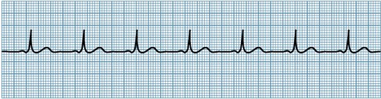
So, a normal sinus rhythm:
http://nuclearcardiologyseminars.com/wp-content/uploads/2013/11/nsr.jpg


  1. Regular (meaning, the number of boxes between each R peak is the same-ish): In this case, about 21 boxes between each peak.
  2. The rate is between 60-100 (remember: 1500/# of small boxes between two consecutive R-R intervals):1500/21=71bpm
  3. You see a distinct P-wave: Yup, little bump right before the QRS.
  4. And the P-R interval is between 0.12-0.2 seconds (3-5 blocks): almost perfectly 3 each time.
  5. And the QRS is tall, and less than or equal to 0.12 (3 blocks) long: almost perfectly 3 each time, also.


Here's a rundown of common dysrrhythmias.

Sinus bradycardia:

  1. regular
  2. rate 1500/# r-r interval boxes <60
  3. P wave present
  4. P-R interval 3-5 blocks
  5. QRS tall, less or equal to 3 blocks
Causes: IICP, hypoglycemia, hypothermia, Valsalva, medications (B-blockers, dig, Ca2+ channel blockers), but it can also be a natural reaction to be really athletic or sleeping, so it's only treated with atropine, dopamine, epinerphine, pacemakers, and the existing medications that are slowing the heartrate should be reduced or discontinued if the patient is symptomatic.

Sinus tachycardia:

  1. regular
  2. rate 1500/# r-r interval boxes >100
  3. P wave present
  4. P-R interval 3-5 blocks
  5. QRS tall, less or equal to 3 blocks
Causes: physiologic/psychological stress such as dehydration or hemorrhage, angina or general pain, fever, etc., and stimulants, or stimulant drugs like epinephrine, norepinerphine, atropine, caffeine, theophylline, hydralazine, and pseudoephedrine.  Treatment: Treat the stressor, vagal maneuvers, B-blockers, adenosine, Ca2+channel blockers, synchronized cardioversion.**

  1. irregular
  2. rate 1500/#r-r interval boxes 60-100
  3. P wave "looks funny," eg biphasic (squiggly looking)
  4. P-R interval 3-5 blocks
  5. QRS tall, less or equal to 3 blocks.
Causes: Stress or fatigue, hypoxia, electrolyte imbalances, hyperthyroidism, COPD, heart disease, valvular disease, or use of caffeine, tobacco, or alcohol.  This may indicate a reentry mechanism that may become SVT, or enhanced atrial automaticity.  Treatment: Beta blockers and cessation of sympathemimetic drugs.  

rate: varies
rhythm: regular atrial flutters up to 350 bpm accompanied by slower ventricular regular or irregular rhythms
P-waves are sharp and irregular, giving a "saw toothed" appearance.
P-R interval not measurable because of multiple P-waves.
QRS usually normal.

Causes: Almost always some cardiac disease (CAD, HTN, mitral valve, PE), cor pulmonale, hyperthyroidism, dig, quinidine, epi, which will cause decreased cardiac output and clotting.  Treatment: Blood thinners (heparin, then warfarin, slow ventricular contractions if needed with Ca2+ Beta blockers, and electrical cardioversion if unstable, otherwise antidysrhythmics (ibutilidem, amiodarone), and radiofrequency catheter ablation.


Rate & Rhythm irregular, very tiny atrial oscillations that are completely unmeasurable (I don't care what your books says, you're not going to be able to count 600 oscillations).  Ventricular contractions irregular
P "fibs"
unmeasurable P-R
QRS usually normal.

Causes: multifocal ectopi due to underlying cardiac disease (even pericarditis, and also surgery), tyrotoxicosis, ETOH, caffeine, electrolyte disturbances.  Treatment: the priority is to decrease HR *if* the ventricular response is "uncontrolled" or "rapid" (>100) with B-blockers, Ca2+ blockers, dig.  Otherwise, if the patient is symptomatic (losing consciousness) cardioversion ONLY IF the patient has be in a-fib for less than 48 hours and/or a transesophageal echocardiogram rules out clots in the atria.  If they have been fibbing for more than 48 hours, they have to be on 4 weeks of warfarin (INR: 2-3) before you cardiovert or you could dislodge a clot and kill him or her.  Non-cardioversion antidysrhythmic options are amiodarone and ibutilide.


Junctional Dysrhythmias: When the SA node fails and the AV node becomes the pacemaker, and the impulse moves backwards, producing a P wave occurring before, after, or under the QRS complex.  P waves appear to be premature atrial contractions.  

Rate: regular
Rhythm: often bradycardia (the AV node beats 40-60), but if some SA node impulses occur, it can be higher when the sympathetic stimulation  to improve CO.
P waves vary, but often are "invisible" as they are covered by the QRS, and sometimes inverted
P-R interval varies
QRS usually normal

Causes: cardiac diseases, digoxin, nicotine, amphetamines, caffeine.  
Tx: atropine for bradycardic rhythms, B-blocks/Ca2+/amiodarone for tachycardic rhythms.

The AV blocks:
1st degree AV block--Normal rate and rhythm for A&V, normal P, P-R>0.2 seconds, normal QRS
2nd degree AV block--Normal and regular A, slower V, multiple P:V ratio, P-R cannot be calculated since there are multiple P waves, widened QRS.  Progresses to complete heart block due to His-Purkinje disease.
3rd degree AV block--P appears abnormal because some will be hidden under QRS--this is because it is complete heart block and the pacemaker that is able to set the ventricular rate is the AV node=severe bradycardia,  multiple P:V ratio, P-R cannot be calculated since there are multiple P waves, wide but consistent.   Hemodynamically very unstable.

Causes: medication (B-block, Ca2+, dig) or cardiac damage/valve disorders.  1st degree has no real treatment except to discontinue medications if the block is medication related, 2nd and 3rd degree are treated with atropine, amiodarone, and pacemakers.

Paroxysmal supraventricular tachycardia: technically any ectopic anywhere above the birfucation of the Bundle of His, usually triggered by an extra pathway that bypasses the AV node causing a reentrant phenomenon (like with Wolff-Parkinson-White syndrome, which is a type of atrioventricular reentrant tachycardia, which is a type of supraventricular tachycardia--yeah, I know, confusing.  You'll get it eventually).  

rate: regular
rhythm: Tachy
P-wave: oftentimes obscured
P-R interval: varies if present at all
QRS: usually normal

Causes: overexertion, stress, deep inspiration, stimulants.  But also from dig, CAD/rheumatic heart disease, cor pulmonale.  Treatment vagal stimulation, adenosine, amiodarone, B-block, Ca2+ block, if hemodynamically unstable, synchronized cardioversion, and radiocatheter ablation.

Premature ventricular contraction: comes from ectopic foci in the ventricles.  If the QRSs originate from different foci, each QRS will look different and that is called "multifocal PVCs."  If they originate from the same foci, the QRSs will look the same and that is called "unifocal PVCs."   Sometimes you have normal sinus, then a PVC--bigeminy.
Sometimes you have 2 normal sinus, then a PVC--trigeminy.
Sometimes you have normal sinus, then two PVCs in a row, then normal sinus--couplet (of PVCs).
If you have 3+ PVCs, that's considered ventricular tachycardia.

rate: anything
rhythm: can be regular, can be irregular, can even be regularly irregular
P-wave: usually hidden under the QRS
P-R: nonexistant (duh ^^)
QRS: wide and distorted

Causes: stimulants, amniophylline, epi, dig, electrolyte imbalance, hypoxia, fever, stress.  Treatment: remove stressor--if hypoxic, give oxygen, if fever, give tylenol, if stress, reduce stress, if stimulants, excrete stimulants, etc.  Give B-block, Ca2+ block, procanimaide, amiodarone.

Ventricular tachycardia: 3+ PVC's.  One type of V-tach is Torsades de Points, which is a polymorphic VT and a prolonged QT interval.  V-tach can quickly degenerate into V-fib.
rate: 250+
rhythm: regular
P-wave: invisible
P-R: nonexistant (duh^^)
QRS: varies

Causes: Prolongued QT (sometimes caused, ironically, by antiarrhythmics), genetics, heart damage (MI/CAD), CNS disorders/electrolyte imbalance.  
Treatment: If pt has a pulse: procainamide, sotolol, amiodarone, implantable cardioverter-defibrillator
If a patient does not have a pulse: CPR, D-fib.  If d-fib unsuccessful, follow with vasopressors (epinephrine), antidysrhythmics (amiodarone).

Ventricular fibrillation: tiny squiggly line.  NO PULSE.  CPR, D-fib.  If d-fib unsuccessful, follow with vasopressors (epinephrine), antidysrhythmics (amiodarone).

Pulseless Electrical Activity:  PEA is where you see electrical activity on the screen, but there is no pulse in the patient.  It is most commonly seen after D-fib (for v-fib or hemodynamically unstable v-tach), and the only thing you can do for this patient is CPR and try to figure out what is causing the PEA.  Causes include: The H's and the T's (see below)
  • hypovolemia (give volume, obviously: PRBCs, D5W or .9NS wide open, elevate the legs, do whatever you need to do to make the systemic volume go up--remember pressors only work if there is adequate volume in the body already)
  • hypoxia (O2, really sucks if it's carbon monoxide poisoning...)
  • met. acidosis (bicarb, regular insulin with D20W, potassium if the etiol is hypokalemia--highly unlikely, I know--and intubate that person because you better believe his ventilatory drive is in the toilet with respiratory fatigue from trying to blow off CO2.)--Yeah, I  know this is neither an H or a T.  Whatever.  It was in my textbook.
  • hypokalemia (give potassium IV DILUTED)
  • hypoglycemia (D20W)
  • hypothermia (warm .9NS)
  • toxins (antidotes, dialysis, chelation)
  • cardiac tamponade (decompress the pericardium--no, not you, the doctor)
  • thrombosis (suck that sucker out--not you, the doctor)
  • tension penumo (insert chest tube)
  • trauma (fix it, whatever it is)
Sudden Cardiac Deathif I remember correctly, usually happens to young people who were just really active sending them into Vtach or V-fib, and you just need to do CPR and D-fib until help arrives with epinephrine and amiodarone.

Prodysrhythmia: this is when antidysrhythmic medication like dig, or amiodarone causes the same dysrhythmias for which they were prescribed.  Yup.  It happens.  I don't know how to treat it directly--that is the doctor's job to figure out.  I just treat according to what the EKG looks like until the doctor figures it out.

Asystole: CPR, treat the cause.  Epinerphine, vasopressin.    Causes include: hypoxia, acidosis, electrolyte imbalance, drugs, hypovolemia, cardiac tamponade, tension pneumo, MI, hypothermia, trauma.

But basically, I hope you started to see a pattern here:  Any time there's an inconsistent rhythm you were given an antiarrhythmic like amiodarone or ibutilidem.  Any time the heart was going too slowly, there was atropine.  Any time the heart was going too fast, there was B-blocks, Ca2+ channel blocks, dig.  Any time the patient was hemodynamically unstable but still had a pulse, cardioversion.  Once there was loss of consciousness but still electrical signal, CPR, then d-fib, epi, and amiodarone--except with PEA, where you have to figure out all the hypos or to toxins, thrombosis, tension pneumo, or trauma.  


CARDIOVASCULAR SYSTEM DISORDERS
btw, Khan Academy (which you should all donate to), has free tutorials and NCLEX-RN questions on this cardiac dysrhythmias.

Quick repost of lab values:
ESR: 1-13 (the Mayo Clinic says 1-22**)
C-reactive protein: risk for CAD  normal <1; average 1-3; high risk >3
PTT: 16-40; aPTT 1.5-2.5X norm
PT/INR: 11-12.5/0.7-1.8 (2-3 on coumadin)
BUN: 10-20          Video on BUN here
Creatinine: 0.6-1.2    
LFTs:
  • Cholesterol <200
  • Triglycerides <150
  • LDL <130
  • HDL <65
  • Albumin: 3.5-5   
  • AST aspartate aminotransferase and ALT alanine aminotransferase 0-35 (signs of hepatitis)
    Video on liver function tests here
  • ALP alkaline phosphatase 30-120 (biliary tree obstruction)

BNP: no failure <100, possible failure <300, failure >300 (300 mild, 600 moderate, 900 severe)
Creatinine phosphokinase MB (CK-MB): <5% (<25 IU/L)
Troponin: <0.2

Here are a few ECG variations you should remember for Acute Coronary Syndrome:
MedSurg by Lewis Dirksen Heitkemper Bucher 9th edition, page 806.
Note, how ischemia (reversible) will result in an ST-segment depression >1box in at least 2 leads, and possible T wave inversion; injury will result in ST-segment elevation; infarction (cell death) causes BOTH the ST-segment elevation, and the T-wave inversion.  Often times if a patient is not showing signs of an acute attack, yet the ECG shows signs of ST-segment elevation or T-wave inversion, you can tell that they had a heart attack in the past.  This is pertinent with cardiovascular disorders.

Hypertension:  Ok, so hypertension can occur because of multiple reasons (pheocromocytoma, renal failure, heart failure, bad diet, and the race most affected is African Americans, especially the men) but whatever the case may be, in the end you need to do the same basic things: eat a heart healthy diet (low sodium, low fat), exercise.  If your hypertension is bad and has caused damage to your kidneys, AND/OR your hypertension is now worsening because your kidneys have conked out on you (remember, you can tell that a person is in renal failure if the creatinine>0.6-1.2; and especially if both Creat:BUN are elevated but the ratio is still 1:10 to 1:20, so like Creat 20:BUN 300.), you need to offload that fluid before you drown (pulmonary edema) with ACE-inhibitors and diuretics, and you need to help your poor, beat up heart (Ca2+ blockers, B-blocks, dig) beat better.

I'll do a review of these medications in another post.

Angina: comes in 3 forms:
1) stable (triggered only by exertion and stress)
2) unstable (fatty deposits rupture/blood clots form, suddenly blocking a narrow artery=suddden decrease in blood flow) that is not relieved by medications
3) variant aka Prinzmetal, very rare, and is a spasm in the coronary artery.

Risks are (of course) family history, smoking, hyperlipidemia (cholesterol>200; Triglyc>150, LDL>130, HDL>65), which often accompanies sedentary lifestyle and obesity, and diabetes.

If a patient presents to the ER in acute onset of angina (s/s of acute coronary syndrome), assess the patient for location of pain, character of pain, duration of pain and whether it was relieved with rest or nitro on scene, and what precipitated the onset of angina (and of course eliminate those triggers to avoid addition attacks while in the ER).  

then, administer MONA in this order:
1) Oxygen (ABC)
2) Nitro
3) Aspirin
4) Morphine
and then, 5) thrombolytics.

and, if necessary, prepare for transport to the cath lab or surgery.  If the patient is stabilized and the doctor feels that it is unnecessary to do PCI, then anticipate beta blockers, aspirin, nitrates, statins, Ca2+ channel blockers, and angiotensin converting, enzymes.

In the real world, you may give MONA immediately without getting an ECG, because the sooner oxygen and blood flow is restore, the more likely you are to recover from ischemia and not progress to injury or infarction, but for the purposes of the NCLEX, you always assess first--it is NCLEX world in perfect harmony, and the patient will not die before you finish your assessment.

Cardiac catheterization: There are two types of cardiac cath for ACS: coronary angioplasty (a balloon is inflated at the site of blockage and mashes the fat against the wall to create a bigger opening), and coronary stent (the same thing as an angioplasty, but you leave behind a fine mesh to keep the fat from collapsing in as soon as you back out of there).

The main things you need to remember as a nurse for cardiac cath is: it's percutaneous (percutaneous coronary intervention), may require dye (check for shellfish allergy--although, many of my doctors have said that the correlation between shellfish allergy and dye allergy is a debunked myth, so who knows), so you will need to:

  • NPO
  • verify consent
  • shave prep (always a trick question whether to shave or to cut hair at the site with scissors to microcuts and prevent infection.  I've never known sources to agree--it's like, whatever, you're going to use betadine/chlorhexadine in a second, who gives a fuck.)
  • explain dye--"you may feel flush and taste a metallic tang," and check for allergies
  • mark baseline pulse

Your main concern is hemorrhage through the femoral artery (and it has to be the artery because you're going into the coronary arteries and that's the only way to them--it's not like you can go through the ICV and thread through the alveoli and back out into the aorta), so:


  • APPLY PRESSURE FOR 15 FULL MINUTES (at least, especially if PT>12.5/INR>2-3), oftentimes you'll use a sand bag to maintain pressure, and KEEP THE LEG EXTENDED--in fact NO HIP FLEXION IN BED for 4-6 hrs.
  • monitor hemorrhage q 15 min for the first 4 hrs, then 30 minutes, then hourly, etc.
  • monitor neurovascular q 15 min for the first 2 hours, then 30 minutes, etc.
  • increase fluids (for hypovolemia, and to wash out dye)
Anticipate the doctor will put the patient on the same medications as for a patient with stable angina without PCI, that is: beta blockers, aspirin, nitrates, statins, Ca2+ channel blockers, and angiotensin converting, enzymes.  You'll have to teach the patient to avoid the valsalva maneuver, avoid stress/sudden bouts of physical activity (ESPECIALLY in the cold--Jesus.  All of those people who suddenly decide that they're going to shovel their house out of a blizzard and end up dying of heart attacks on their sidewalk), rest after eating (especially hard to digest things, like steak), and stop smoking.  Exercise LIGHTLY--seriously, walking is enough--and eat a heart healthy diet.  (Like, duh.)

So, with the medications you have to teach:
nitro--SIT YOUR ASS DOWN, take with onset of angina SL q 5 minutes up to 3 times.  Call 911 if it's not relieved after the first tablet.  Keep the nitro in a dark, dry container out of the light and away from heat, and replace q 6 months even if you've never used it--don't be cheap about this medication.  If you're taking it and you don't get a headache, maybe the nitro isn't working.  If you're taking nitro on a daily basis (as a cream or a patch) you have to use it for ONLY 12 hours a day (applying it in the morning) and wash it off for the other 12 hours to prevent developing a tolerance.  And do not take it if you are on a sildenafil (Viagra) because your blood pressure will bottom out and you will die.  I mean, be nicer than that, but make sure that the patient gets that this is serious.

Ok, so you've had angina, and a cardiac cath, and you're still eating McDonalds, or you haven't given up your job as a top litigator and then you decided to go shovel your house out in the middle of a blizzard and you've had a full on grade A heart attack and given yourself a massive myocardial infarction.  Maybe you went into V-fib, had to be D-fibbed and had someone pound on your chest and break a few ribs the whole way from your house in the back of an ambulance to the hospital, where they discovered that a piece of artherosclerotic gunk broke off in spite of the coronary stent, and got lodged in your left coronary artery.  They cracked you open, did a CABG on you, and now you're in ICU, in extreme pain (fun fact: diabetics, due to diabetic neuropathy, may not report pain), and your ECG is showing simultaneous ST-segment elevations and T wave inversion.  (Which reminds me, I really have got to exercise today...)

Myocardial infarction:  s/s: severe chest pain (or not, with diabetes, women or elder patients) unrelieved with nitro or rest that crushes and radiates to jaw, left arm, neck, back, etc.  Diaphoresis, N/V anxiety, ECG shows tachycardia/dysrhythmias c possible ST elevation (STEMI) and/or T inversion, hypotension + dyspnea.  Expect troponin>0.2, CK-MB>3,  LDH (lactic acid dehydrogenase) >140-200, 

Nursing goal: Rest the myocardium and preserve heart muscle.

MONA: 
Immediately: Oxygen, nitro, aspirin, morphine, streptokinase or alteplase (within 6 hours), amiodarone, calcium channel blockers.
Later: stool softeners + bedside commode, promote self care and exercise but stop if fatigued or angina occurs, heart healthy diet, stress management, smoking cessation.
Discharge medication: heparin, aspirin, warfarin, enoxaparin, clopidogrel (antiplatelet), metoprolo, diltiazem (Ca2+), nitro, simvastatin (antilipidemia).

The result of a myocardial infarction is heart failure.

Heart failure:  A chronic condition where the heart is unable to meet the body's oxygen requirements, and is often accompanied by fluid overload/inadequate tissue perfusion + fluid retention due to decreased renal circulation.  Depending on what caused the heart failure (cor pulmonale, which coronary artery was blocked, kidney failure, hypertension, etc.) there may be L.S. HF (symptoms are related to decreased systemic/pulmonary circulation, i.e. elevated PAWP, pulomonary edema and paroxysmal nocturnal dyspnea, low O2 sat), or R.S. HF (symptoms related to systemic congestion, i.e.  acites, hepatosplenomegaly, dependent edema + fluid weight gain, elevated CVP, JVD, anorexia/vomiting)

Ok, wait, but what is the PAWP and the CVP?  I ask, because I know it has to do with catheters measuring pulmonary artery wedge pressure (PAWP) and central venous pressure (CVP), but that's about all I remember.  So, let's take a side trip into HEMODYNAMIC MONITORING.  If you already got the theory, here's the hemodynamic values you should know:

CVP: 1-8mmHg
PAWP: 4-12 mmHg
Cardiac output 4-8L/min = Stroke volume x Heart Rate
Mean arterial pressure 70-105 mmHg = [systolic + 2diastolic]/ 3
Systemic vascular resistance 1970-2390 gynes/sec/cm^-5= [(MAP-CVP) x 80]/ CO
Arterial hemoglobin saturation 95-100%
Mixed venous hemoglobin (SCVO2): 60-80%

If you didn't take the side trip, basically, you need to know that elevated levels of CVP (>8)and PAWP (>12) mean that the heart is (for whatever reason) unable to pump blood efficiently.  In the case of heart failure, it's because of heart failure (duh).

Treatment of heart failure= pump it, park it, pee it=focuses on decreasing fluid volume overload and helping the heart to pump more efficiently (inotropic medications)/decreasing systemic vascular resistance.  
Medication for inotropic (pump it): digoxin
Medications for decreasing systemic vascular resistance (park it): B-block, Ca2+ ch. block, nitrates/hydralazine, nesiritide, milrinonedobutamine.
Medications for decreasing fluid (pee it): ACE inhib, ARB, diuretics


Coronary artery bypass graft (CABG): usually requires an open chest surgery with bypass (ECMO).  Your job as a nurse is to:
preoperatively: VS, assessment, HX.  support (psych and anxiolytics), teach the family and patient to anticipate intubation, IV lines, foley, arterial line, chest drainage tubes.
postoperatively: hourly assessement for first 8 hours of complications:
  • vitals, neurological (pupils, reflexes, Glasgow)
  • cardiac: CVP, PAWP, ECG, sounds, increased bruising, etc., peripherals
  • resp: ventilator settings
  • renal: I&O, electrolytes, urine specific gravity
  • pain: location, type, intensity, onset
DVT: usually caused by increase in thrombi formation (think estrogen in pregnancy or contraceptives, or long term immobility from injury, immobility, or age).  You'll see a red, hot swelling of one limb, that may be bumpy, knotty, and shiny (edema leaking through).  You may see a venous ulcer (usually medial side of the leg at the ankle or above).  It is best prevented by ambulation, weight loss, wearing supportive antiembolitic socks, and elevating the lower extremities and not crossing your legs, and preventative SQ heparin.  It is diagnosed with an MRI, Spiral CT, U/S, and then treated with heparin (PTT 60-100) and warfarin (INR 2-3), alteplase, elevating the extremity, applying warm, moist compress, and being vigilant for s/s of PE, stroke, or MI.  Otherwise, sclerotherapy, ligation and stripping, or thermal ablation may be necessary.  

Peripheral artery (occlusive) disease (PAD): Peripheral arterial disease is just a buildup and consequential narrowing of arteries.  It is a significant risk for future major coronary events--note the name "peripheral artery disease" is misleading because atherosclerosis can happen all over the body, such as in the heart (CAD) or in the brain (cerebrovascular disease).  As such, the risk factors of PAD is the same as those for CAD, which are, high cholsterol >200, high triglycerides >150, and elevated CRP >3, smoking, diabetes, etc.  S/s include: intermittent peripheral claudication pain that resolves with rest (because decreased blood flow will exacerbate the removal of lactic acid), and calf muscle atrophy, and shiny skin with hair loss, paresthesia, and thick toenails (signs of decreased oxygen and nutrients reaching cells), and may lead to leg ulcers.

Leg ulcers are another thing that I found really confusing because it's never really well explained.  I'm still not good at it, but as I've said before, I've accepted that there are many things that will make sense to me, and that I will learn after I've passed the NCLEX.  To see a quick differentiation of leg ulcers, take a look here.  

Along the same vein, there is a similar disease called...
Thromboangiitis obliterans aka "Buerger's disease": a disease that causes repeated inflammation of the blood vessels of the extremities, resulting in idiopathic thrombosis and occlusion.  This also has the symptom of intermittent claudication, inflammation due to clots, and cold skin that is relieved by rest, smoking cessation, avoiding caffeine, and avoiding constrictive clothing or the cold.  If it is prolonged open sores may occur on the fingers and toes that require amputation.

To make things more confusing, there is another similar disease (that actually popped up on my NCLEX-PN as the final question) called...
Raynaud's syndrome: a disease that causes vasospasms/constriction of the arteries due to exposure to cold and stress.  These may lead to ulcerations of the fingertip, so the patient should wear extra clothes in the cold, but make sure that the clothes are not restrictive.  Smoking cessation, and limiting caffeine intake is also suggested.  You could also administer nifedipine.  Nifedipine would NOT work with thromboangiitis obliterans because there is inflammation and thrombosis involved.


Structural Cardiac Disorders
Valvular disorders: valve disorders are mostly cases of stenosis which makes it difficult for the heart to eject blood, but also, allows for backflow (leaking) back into the chamber that was just pumping.  Just remember the side each valve is located (R: tricuspid, R pulmonary, AV; L: mitral, aortic, AV).  Murmurs are just the turbulence of blood sloshing around erratically because of the stenosis or prolapse.  Mostly it leads to heart failure.  The only treatment is surgery for replacement, which will require warfarin for life if it's mechanical, and immune suppressants if biological (but warfarin only for 3 months).  If you have an autologous fancy, valve, you won't need either except in the immediate postoperative.  All of the valve surgeries will require prophylactic antibiotics for any further procedure to prevent infective endocarditis.  

Aortic aneurysm: usually caused by congenital connective tissue disorders, but also from infection and atherosclerosis.  An aortic aneurysm greater than 5 cm requires surgical intervention.  Usually a patient doesn't even know he has an aortic aneurysm.  He may complain of:
  • thoracic pain, dyspnea, hoarseness, cough, and dysphagia (remember, the larynx, pharynx, esophagus, trachea, are all in the mediastinum with the aorta and the heart)
  • lower aneurysms (remember, the abdominal aorta is still part of the aorta) may cause abdominal pain, persistent or intermittent low back or flank pain, and you might actually be able to see a pulsating abdominal mass
Your job as the nurse is to: 
Preoperative: maintain systolic between 100-120 with beta-blockers, antihypertensives.  Continuous nipride might be prescribed, and to be vigilant for signs of rupture (sudden, intense pain with rapid BP drop).  Also, you'll have to care for the patient during CT scans, MRIs, X-ray, and ultrasound.
Postoperative--basically the same as preoperative because now, instead of an aneurysm, you've got a line of sutures holding the aorta together.  Freaking hell!  And of course, infection.


SHOCK

Ah, shock.  So many versions.  So confusing.  I'd encourage you to make a chart in the end to get an overview, but I'm just going to talk about each separately.  I personally found this Khan Academy video very helpful.  There is also a collection of review questions from Khan Academy.  As I said before, I highly suggest donating to them since they provide excellent education for free, which helps those who come from socioeconomic hardship to have the same education someone of means might get from a private school.  Here's a great story showing that.  

First off, shock is any situation where there is decreased tissue perfusion and impaired cellular metabolism, resulting in cell starvation.  There are 4 main categories: cardiogenic, obstructive, hypovolemic, and distributive.  

Cardiogenic shock, obstructive shock, and hypovolemic shock are the ones you'll most see on TV. They're slightly difficult to tell apart because they all have some form of decreased cardiac output, and in response the blood vessels constrict in attempt to increase blood return to the heart.  In addition, all three have decreased mix oxygen content (MVO2) because the body's cells are desperately trying to pull as much oxygen out as possible, resulting in a low oxygen content in the blood returning to the heart.  They deviate when it comes to preload levels.  In cardiogenic and obstructive shock, you'll see an increased PAWP>12 (remember, this measures how much pressure is in the ventricles at the end of diastole) because the ventricles are unable to properly eject blood out of the heart either because the heart is damaged (like from an MI) and unable to pump (cardiogenic shock), or because there's an obstruction (like an aortic stenosis or cardiac tamponade) preventing the blood from flowing (obstructive shock).  In hypovolemic shock, there simply isn't enough volume in the body to properly fill the ventricles (because of dehydration or hemorrhage) resulting in a lower PAWP<4.  With cardiogenic shock you have to fix whatever damage there is to reestablish proper circulation, and with obstructive shock you have to either unplug the thrombosis or open the stenosis.  With hypovolemic shock you have to fix the source of the bleed, or simply refill the body with blood components and/or fluid.  The emergent steps to stabilize a patient (for surgery): 1) Establish airway, give O2, and 2) improve circulation and contractility with epinerphine (vasopressor) and IVF, and 3) then repairing the problem.  

Distributive shock: Neurogenic, septic, and anaphylactic
Neurogenic shock, which occurs when the spine is injured, causing an impairment in sympathetic response.  Neurogenic shock is the only shock that results in a BRADYCARDIA and simultaneous decrease in nervous system tone (blood vessel dilation).  So, the spine is injured, HR drops, causing cardiac output to decrease (SV x HR= CO<4L/min), and the blood vessels aren't constricting.  PAWP isn't really affected even though there is decreased vascular resistance. Mixed oxygen saturation should be low because the blood is traveling slower so the surrounding tissue has more time to absorb O2.  Treatment should be to maintain BP: pressors, IVF, atropine (blocks the parasympathetic nervous system=stops rest and relax signals to increase the heartrate).  

___________Detour into neuro for a second:  SCI

With SCI, if the injury is above T6, there is a risk of autonomic dysreflexia should the patient experience bowel impaction, bladder distension, triggering of pressure points, ulcers, or pain.  Autonomic dysreflexia is a life-threatening syndrome of sudden, extreme hypertension, with bradycardia (30-40bpm).  Other s/s: H/A, piloerection, flushing above the level of the injury, blurry vision, nasal congestion, and nausea.

The order of nursing actions is:

  • High fowlers (I know, your first instinct is to roll that person over and try to un-impaction him, but, don't, cause that may not be the problem.  Putting the person in high fowlers buys you time to assess the causation)
  • (have someone else) alert the physician
  • determine and remove causative stimuli--also remove anything that may cause more stimuli to the patient
  • give medications (lidocaine, to help with digital fecal removal and/or bladder cathing, a-blockers, and Ca2+ channel blockers)
  • teach bowel and bladder management

________________________End detour


Septic shock and anaphylactic shock are another couple that have strong similarities.  Septic shock and anaphylactic shock both have immune mediated systemic vasodilation, which causes edema (resulting in decreased circulating volume, and makes it more difficult for O2 to diffuse from the blood vessels to the tissues). In both, PAWP is marginally decreased, but not by much, if at all.  The treatment for septic shock is 1) (Airway, Breathing) ventilation; 2) (circulation) IVF, pressors, 3) treat the source with blood cultures and ABX (desire CRP<1, ESR <20-25 = decreased inflammation, and decrease bacterial count), and the treatment for anaphylactic shock is 1) establish airway with ventilation, 2) maintain BP with epinephrine (and open airway) and IVF, and 3) continue to reverse the allergic reaction with benadryl.


There's also something called dissociative shock in which hemoglobin doesn't release O2.  There are 2 causes of dissociative shock.  The first is: sometimes a person has red blood cells where the iron in the hemoglobin is Fe3+ instead of Fe2+.  Fe3+ (ferric ion=methemoglobinemia) causes Fe2+ hemes to increase in ferrous ion oxygen binding.  This is caused by nitrates, especially antibiotics, (such as TMP/SMX, and dapsone), or anesthetic (like benzocaine), or pesticides.  Neonates are especially vulnerable to methemoglobinemia because they're missing an enzyme that converts Fe3+ to Fe2+.  Treatment of methemoglobinemia is IV methylene blue, which causes Fe3+ to be converted back to Fe2+.  The other cause of dissociative shock is CO (carbon monoxide) poisoning which has a 100X greater binding ability to hemoglobin than O2.  It causes a change in shape of the hemoglobin, preventing their release.  The cause of CO is carbon combustion, like a fire burning stove.  Treatment is oversaturation with O2 in a barometric chamber.


Wednesday, January 13, 2016

Day 13-12: Nursing Care of Children--Newborn and Infant Standard Tests, Complications and Congenital Anomalies

Oh, newborns.  So exciting and cute.  So freaking complicated and scary.  I mean, all of nursing is complicated and scary, but babies, especially.  Because they're small.  And fragile.  And weird.  Their circulation is weird, and their breathing is weird, their vital signs are weird.  They're wet.  And slippery.  And floppy.  And you have to poke and prod and test their Moro reflex, which makes them cry, and you just about lose your shit because evolution has wired us to want to give babies whatever they want to make them stop crying.  

Before we get into APGARS (yay, more to memorize...)

Newborn VS:

HR: <100 if sleeping, 110-160, up to 180 when crying.  Protip: I find tapping my finger while counting their tiny apical pulses for a full minute helpful with babies because otherwise I lose count.
Respirations: 40-60, usually shallow and irregular.  There is transient apnea, but the apnea should never be more than 20 seconds.  Look for nasal flaring, intercostal/supra/subcostal retractions, or see-saw breathing.  
Temperature: 99.7-98.9F axillary.  Temperature instability is a sign of infection.
BP: 60-80/40-50.  Hypotension=sepsis/hypovolemia; Hypertension coarctation of aorta.  


So, you delivered the head, cleared the mouth, then the nose (the mouth first to prevent aspiration), delivered the rest of the body, cut the cord, now what?

Apgar was named after the person who invented it (Virginia Apgar), yes, but more importantly, APGAR stands for:
A: Appearance completely blue (0); acrocyanosis (1); completely pink (2)
P: Pulse zero (0); <100 (1); >100 (2)
G:Grimace (with stimulation) no response (0); grimace (1); crying (2)
A: Activity (is the baby flexed) limp (0); some flexion (1); well flexed (2)
R: Respiration: zero (0), weak/<40 (1); good cry/>40 (2)

The numbers you need to remember with APGAR are: 4 and 8.  4 (repeat 4, 4, 4, 4, 4, 4) and up is moderately distressed (3 and down is severely distressed), 8 and up is WNL.  APGARS are taken at 1 minute and 5 minutes after birth, and only again at 10 minutes if the child does not improve/is still below 7 at 5 minutes.  Note, the stimulation for grimace is just drying the baby and getting him warm.  


Once that is complete, the baby is printed and secured with an identity band for mom, dad (or mom mom or dad dad or whatever), and baby.  And as long as baby is stable, we can begin feeding immediately after birth, and every 2-3 hours after with breastfeeding, and 3-4 hours with formula (formula digests slower, and you have to burp them ever 1/2 ounce or 1 ounce, and you have to chuck it after 1 hour of being prepared to prevent bacterial contamination).  Here is proper latch on positioning, note the football position for C-section moms.  We want a minimum of 100mL/kg in the first 24 hours.  So, for example a 6 pound baby would be 2.7 kg x 100 mL/kg=270 mL.

[FYI: 16 ounces/1lb
so,  if the baby is 6lbs 7 ounces 
that's 6pounds + (7ounces/16ounces in a pound)= 6pounds+ 0.43pounds=6.43 pounds
--> convert to kilos
6.43pounds/2.2pounds per kilo=2.9kilos
--> multiply that times 100 mL/kg in the first 24 hours
=290 mL
but if they give you a problem, then the creator of that question is an asshole.
But you might have to calculate that in real life.]

GU: the baby needs to pee within the first 24 hours of life, and after the 4th day of life, urination occurs 6-10 times a day.  They usually poop within 48 hours.  Poop usually looks yellow and seedy. Teach that circumcised penises can be cleaned with warm water, and to apply petroleum jelly with each diaper change (q 4 hours minimum), and that yellow mucus is ok and to leave it on.  It should heal in 2 weeks.  Don't submerge the kid in a bath until then, cause the umbilical cord has to dry and fall off first (and you just clean that with a neutral pH cleanser and sterile water at birth, but otherwise it should remain dry until it falls off).  


For the Ballard Score go here.  I'm not going to memorize that because I don't have time for that.

Standard care and labs for newborns include:
  1. eye prophylaxis with erythromycin (for GCCT--aka gonorrhea and chlamydia) within the first hour of birth
  2. vit K IM injection within the first hour of birth
  3. Hep B within the first 12 hours.  
  4. ABO Rh type and screen
  5. PKU
  6. CBC:
  • RBC 4.8-7.1 million
  • Hgb 14-24, Hct 44-64
  • WBC 9-30,000
  • platelets 150,000-300,000
  • glucose 40-60  (within 2 hours of birth)**
  • bilirubin day 1: 0-6; day 2: <8; day 3 <12;  hyperbilirubinemia >15 at any time 
Any time a baby is stressed (from withdrawal, from cold, from illness, from RDS) it will burn more calories.  It is important to check glucose (hypoglycemia=2 consecutive <40 in term, <25 in preterm--remember preterm is before 36 6/7 weeks).  Babies with hypoglycemia are jittery, cold and clammy, and eventually become flaccid and/or have seizures and slip into a coma--AND IT HAPPENS QUICKLY.

Transitional Periods
After the baby is born, it has 3 transitional periods: 1) first period of reactivity=first 30 minutes of life, which is the best time to breastfeed, 2) sleep from 30 minutes to 2 hours old, 3) second period of reactivity from 2 hours till 8 hours, where gagging and vomiting usually occurs, and then they feed again.  (Awww, so cute.)


Guthrie test:
http://www.homebirth.net.au/wp-content/uploads/2010/02/new+born+screening-450x237.jpg

Phenylketonuria
Every baby is tested for phenylketonuria 2 days after birth by federal mandate, with the Guthrie test (heel stick) which is a genetic condition where the baby cannot break down phenylalanine from protein into tyrosine.  The infant starts off with failure to thrive, vomiting, irritability, hyperactive/erratic behavior and ends up with brain damage (older children display schizoid behavior like catatonia, screaming, biting, and head banging).  Treatment means a protein restricted diet with no sweetners for life, and the baby must have a special phenylalanine-free formula (like Phenex-1).  


Substance Dependent Babies
Fetal ETOH syndrome: Since ETOH is a depressant, the lack of a depressant will result in an irritable baby, crying, seizures.  Other problems are IUGR, smaller/thinner/flatter than normal craniofacial features, and possible deafness.
Nicotine: premature, low birthweight, increased COPD (bronchitis, pneumonia), developmental delays, and increased risk of SIDS.
Opioid: sneezing, poor temperature regulation, hyperactive Moro reflex, CNS irritability-->seizures (especially with methadone).
Cocaine: shrill cry, tremors, sleeplessness, muscles spasms, feeding problems.

Often phenobarbital is prescribed for seizures.  Decrease stimuli, swaddle, small frequent feedings of high calorie formula (possibly gavage).  Many struggle with poor sucking of they can settle down enough to eat at all, so preterm nipples with larger holes are used, but try to breastfeed because it has been found to help ease withdrawal symptoms (the drugs carry over, especially if mom is on methadone to help kick the habit).  


Premature Babies (<38 wks)
Premature babies have a low birth weight, little fat (brown fat deposits insufficient, which increases impact of cold stress or oxygen deprivation stress so cluster care and keep the baby warm), long periods of apnea (10-15 seconds), and may not be able to suck/gag/swallow/cough (before 34 weeks) so will require parenteral nutrition.  Also, the child may suffer from RDS.

Respiratory Distress Syndrome
Occurs with premies, babies with meconium aspiration (aka perinatal asphyxia or stress asphyxia), and with C-section babies because they haven't had enough surfactant produced (premies), or they have fluid in their lungs (part of the compression of the torso required to pass through the pelvis squeezes out the fluid from the lungs and helps the baby draw its first breath).  Oftentimes they will have intercostal or substernal retractions, nasal flaring, grunting, rales, cyanosis, etc.  So, administer beractant (a synthetic surfactant), and don't suction for 1 hour so it can spread across the inside of the lungs.

Post-term Babies (>42weeks)
Post-term babies are at risk for injury during delivery (shoulder dysotcia, fx clavical, brachial plexus injury, or facial paralysis), possible risk of degenerating placenta causing meconium aspiration, hypoglycemia, and jaundice.  Initiate early feeding, and watch for signs of pneumonia or jaundice.

Hyperbilirubinemia 
(Bilirubin >15)
There are two types of jaundice: physiologic ("natural" or "expected") and pathological (has a disease process).  Physiological jaundice occurs gradually (>24 hours later), and is caused by the natural breakdown of extra RBCs from mom.  Also, it can happen because newborns have immature livers (less clotting factors), and there is a lack of Vit K.  Pathological is jaundice that sets in within 24 hours of birth and/or lasts more than 7 days, and has an increase in bilirubin at 5mg or more a day, and is caused by blood incompatibility between the mother and the child, or from an infection, which can lead to kernicterus (brain damage due to bilirubin >25).  Direct Coombs to test for sensitized RBCs.  Treat with phototherapy (break q4hr to unmask eyes and check for inflammation or injury)--keep the kid naked, except for the genitals, and protect from burns (no oils or lotions), dehydration (lots of water), and hyperthermia.  Feed the baby frequently to excrete bilirubin in stools (will look loose and green).  If the baby is at risk for kernicterus (bilirubin >25), you may have to give an exchange transfusion.  Kernicterus starts with the same stress symptoms as any other disease (lethargy, poor feeding, temperature instability, and hypotonia-->progresses to fever, seizures, and high pitched cry, and will result in cerebral palsy, mental retardation, upward gaze paresis, and hearing loss.

An easy way to remember risk factors for hyperbilirubinemia in full term infants is the acronym 
Jaundice <24 hrs after birth (physiological)
A sibling who was jaundiced as a neonate
Unrecognized hemolysis (incompatibility--remember, Rhogam @28 weeks and at birth or any trauma), or delayed cord clamping=increased blood volume
Nonoptimal sucking (slow GI=re-unconjugation of bili in the intestine goes back into the bloodstream)
Deficiency in glucose-6-phosphate dehydrogenase
Infection
Cephalohematomas/bruising during delivery
East Asian or Mediterranean descent


CONGENITAL ANOMALIES: CARDIAC

http://cardiovascularsystemud.weebly.com/uploads/8/2/1/2/8212050/5709166.jpg?822
Quick A&P review, basically the fetus gets oxygenated blood from mom (the umbilical vein--remember vein means blood returning to the *fetal* heart) via the ductus venosus (vein, get it?), where it mixes with the deoxygenized blood from the fetus' own vena cavas.  The fetal heart circulation bypasses the lungs (shunts a little to help the lungs develop, but for the most part nothing goes to the lungs), by 1) flowing through the foramen ovale (a hole between the atriums) to be ejected by the left ventricle into the aorta, and 2) being shunted from the pulmonary artery through the ductus arteriosus (artery, get it?) into the aorta.  Once the baby is born, the foramen ovale and the ductus arteriosus should close, and the lungs open, and the baby gets its own 100% super oxygenated blood in through the left side of the heart just like we adults do.  All circulatory congenital defects have a breakdown in that transition.

Almost all require surgical intervention except maybe PDA or ASD.  These can be fixed with indomethacin which closes the holes.  If left unfixed they all result in poor growth and development, and some degree of heart failure, and some of them (like TGV) result in death.  Atrioventricular septal defects will result in a systolic murmur.

PDA
http://www.drugs.com/mcd/images/image_popup/DS00631_IM01560_r7_patentductusthu_jpg.jpg

PDA--the ductus arteriosus doesn't close.  Most of the time it is asymptomatic, but sometimes there is a heart murmur and a wide pulse pressure because the aorta is getting a fuckload of blood from both ventricles, resulting in a high systolic.  The left ventricle has to work super hard.  Treatment: indomethacin and/or surgery.

Atrial Septal Defect (Patent Foramen Ovale)--The foramen ovale didn't close.  The blood is bypassing the lungs.   Treatment is surgery during preschool years (3-6 y-o).

Ventricular Septal Defect--Really large defects are called "Eisenmenger's complex."  You'll hear a big systolic murmur, and heart failure.  Small defects close spontaneously, large ones require surgery during preschool years.

http://www.drugs.com/mcd/images/image_popup/DS00615_DS00998_IM00438_r7_tetralogythu_jpg.jpg

Tetrology of Fallot--1) Pulmonary stenosis, 2) VSD, 3) overriding aorta, 4) R. ventricle hypertrophy.  This kid is seriously sick, and gets acute cyanosis and hypoxia called "tet" spells.  Squatting helps to increase systemic vascular resistance, reversing the shunting and getting blood to the lungs and helping to oxygenated the blood.  If this is hard to understand, put your finger over the aortic arch and imagine no blood can flow out that way (this is what happens with systemic vascular resistance).  Now the blood has no where else to go when the ventricles pump *but* to the lungs.  This kid usually has clubbing of the fingers and severe growth retardation.  Surgery is necessary within the first few years of life.

TGV with ASD
http://static.bandbacktogether.com/media/images/2012/02/tgvdiagram.jpg

Transposition of Great Vessels--This is the condition I personally call "I don't know how this baby survives 3 minutes after birth," because the aorta is connected to the right side of the heart, and the pulmonary artery is connected to the left side of the heart.  The only way this baby survives is if it ALSO has VSD, ASD, or PDA, or SOMETHING that allows it to somehow circulate oxygenated blood between the two sides.  It's insane.  The treatment is to continue giving prostaglandins to keep the ductus arteriosus open and surgery is immediately done to swap the aorta and pulmonary artery back into the right place.  It's absolutely insane.

https://blogger.googleusercontent.com/img/b/R29vZ2xl/AVvXsEgDG40D8hE0ANywVcGt_I8LIpM_Oym-ChVDMRGpz7w2j7A36g2mUG_AcIluUzO0LcIHAFNCKbOgn0rqvKy0SzyprrPlc9k2FphMqKT7Z9vYPexFx0xZvBZ32nfP7q3T1-zmx4PE-3WD6jLV/s1600/Postductal+coarctation+of+the+aorta.png

Coarctation of aorta--So basically the aorta is narrowed, and the lower extremities don't get enough oxygen or nutrition.  The left ventricle has to work extra hard to fight past that narrowing, causing heart failure.  However, look at what those three little arteries at the top of the aortic arch flow to:

https://www.netterimages.com/images/vpv/000/000/013/13922-0550x0475.jpg
Increased blood pressure to the carotids can cause hypertension with headaches, epistaxis, and intraventricular hemorrhage--remember the infant's blood pressure is only 60-80/40-50 because those little brain blood vessels are very fragile.


The nursing considerations for children with these congenital heart defects are: cluster care, provide small, frequent, high calorie meals with plenty of time to eat (gavage if necessary), avoid making the babies cry (I don't know how to not make a baby cry with gavage...).  Observe for infection since these kids are at higher risk.  Give medications to decrease the labor on the heart--like digoxin (0.5-0.8) hold if AP <90 (this is different than adults where the threshold is 60), Ca2+ channel blockers like captopril or enalapril, and duretics like furosemid or chlorothizide.  


CONGENITAL ANOMALIES: SKELTAL

Clubfoot--To call clubfoot a purely congenital anomaly is a lie.  The truth is we have no idea what causes it.  Probably some sliding scale of genetics to intrauterine positioning.  Club foot has 5 different manifestations you need to be concerned with:

http://odlarmed.com/wp-content/uploads/2009/04/talipes_clubfoot.jpg

1) talipes equinovarus--the most common, with plantar flexion and feed bending midline.  To help you remember the name, equin/o/varus means "the heel elevated like a horse" (hence "equine") and "turned inward" (hence "varus").


http://www.nursing-help.com/wp-content/uploads/2011/08/image60.png

2) talipes calcaneus--the heel down and the toes up as if in permanent dorsiflexion.
3) talipes equinus--again, with the heel elevated like a horse.

http://www.nursing-help.com/wp-content/uploads/2011/08/image59.png


4) talipes varus--as before, the toes turning midline
5) talipes valgus--the toes turning laterally

Whatever the cause, it is identified either with ultrasound or at birth. with radiography done to see the extent of the deformity.  After the child is born, massage for lesser degree malformation can be used to manually straighten the foot/feet, or serial casting (changing the cast q2 weeks for 12 weeks) until maximum correction is achieved, and then application of a (rather medieval looking) Denis Browne splint or reverse last corrective shoes to maintain the correction.  If the correction is too late, or not effective enough, surgery with ORIF may be required between 3-12 months of age.

Danis Browne splint:
https://upload.wikimedia.org/wikipedia/commons/8/83/Botas.JPG
Hip Dysplasia: hip dysplasia, in its most basic form, is when the femoral head and the acetabulum are unstable or dislocated. This can happen with acetabular dysplasia (the abnormal development in utero), a complete dislocation (during breech birth, or LGA, or late-term babies), or subluxation (a partial dislocation)--keep in mind even those that are due to "injury during birth" may also have some genetic predisposition. (Also, remember that during the pregnancy, the mother is releasing progesterone, and progesterone makes everything squishy, including the baby's ligaments.  And the baby itself is cartilaginous.)  This is why after the baby is born we do a physical where one of the jobs is to flip the baby over and look for symmetry of their gluteal and thigh fat folds.  And we hold them upright to see if one knee hangs lower than the other (remember when something is dislocated, the muscles suck the bones past each other so the dislocation actually looks shorter), which is called the "Allis sign."  We also use Barlow (intentionally testing if the femoral head can be dislocated) and Ortolani's (if dislocated, then reducing the dislocation) test.


Yeah.  I know.  Scary as fuck.

The treatment for hip dysplasia is a Pavlik harness that prevents hip extension or ADduction for infants <6 mos, and Bryant's traction and surgery to reduce with infants >6mos (or the Pavlik harness didn't work) with the addition of a hip spica cast.

So the basic care of both hip spicas and long leg casts (in the case of genu varum/valgus, or clubfoot), is ABC (checks q15) and by not applying the cast too tightly or smushing it while its drying, infection/skin care from pressure sores, etc. (you can apply "petals" of padding with tape around the edges of the plaster--especially around the window where urine and defecation take place), and also pain and psychosocial distractions for the baby, and nutritional needs (constipation from reduced movement).  The parent may also need special strollers or car seats.


CONGENITAL ANOMALIES: NEUROLOGICAL

***Remember that AFP, or MSAFP is something you *screen* for in the first trimester with an integrated first trimester screen along with Tri21 and Tri18, and in the second trimester with quad and penta screens.  Normal AFP is <2.5 MoM for singlets.

Neural tube defects--There are three main levels of neural tube defects you need to be concerned with: occulta, which is where everything is still sealed, meningocele, which is where the meninges is still intact and there's just a little bubble protruding out of the back, and myelomeningocele or "spina bifida," which is when there is an open spinal defect and the meninges (remember the meninges are the three layers of spinal membrane covering the nerves that encase the spine and the brain into one sac) are open and the spinal fluid and nerves are all spilling out.

Complications are hydrocephalus, clubfoot, scoliosis, congenital hip dysplasia, bowel and bladder problems...

Treatment is prevention (all women of child bearing years should take folic acid supplements since the spine fuses by week 10 and often times people don't know they are pregnant until after 10 weeks). After the child is born the treatment is surgery.

The nurse's job presurgery is to preserve the integrity of the meninges by keeping the infant prone and covering the protrusion with sterile saline moistened gauze.  You don't cover the sac with clothes or diapers, and you have to be careful to clean the perineal area well to prevent tears or cracks in the meninges or the development of meningitis.  Note that children with NTDs have an increased risk for latex allergy.  Postsurgery, keep the infant in prone, monitor for IICP and hydrocephalus.  If the child's injury causes bladder and bowel nerve damage, teach the parents CRede's maneuver and the child how to self-catheterize around age 5-6.  Oftentimes the patient won't be able to feel if she has a UTI, so it's important to teach monitoring for infection (foul smell, cloudy urine, temperature).

Hydrocephalus-the imbalance of absorption of production of CSF inside of the cranial cavity, and/or the obstruction of the flow of CSF through the ventricular system.  Sometimes, hydrocephalus develops later in life due to a malformation such as a neoplasm, infection, or trauma.

The infant will have increased head circumference with bulging anterior fontanels and distended scalp veins.  They will be irritable, have a high pitched scream, and it is extremely uncomfortable for them to be cradled horizontally.  Children with hydrocephalus like to be held upright--this relieves pressure from their cranium.  An older child may verbalize headaches, blurry vision or diplopia, vomiting in the morning, and even experience seizures.

This kid needs seizures precautions both pre and post-op for realz, and constant neurological checks (like we said before, for children, it is measuring thee circumference of their occiptal frontal region).  After the surgery you position the baby on the non-shunt/non-surgery side (you can use a wedge to keep them tilted up on one side), and watch their VS. like a hawk.  Watch for sudden changes in consciousness, vomiting, and sudden increase in difficulty with feeding.  Also, for infection--cause, you know, meningitis is bad.  Also, teach the family TO WASH THEIR DAMN HANDS when handling the kid especially when pumping the shunt to relieve CSF pressure.

Cerebral Palsy-at it's most basic qualification, cerebral palsy is abnormal muscle tone and lack of coordination with spasticity (often with contractions) either of diplegia (both legs), hemiplegia (one side of the body), or quadriplegia (all 4 limbs).  These children often have delayed milestone development and have a lot of back arching and dystonia or ataxia, but little other spontaneous movement including sucking and swallowing (so feeding can be challenging).  Often these children have intellectual, visual, hearing, speech and language deficits, and can have seizures.  Most cases of cerebral palsy are due to brain insult, whether in utero, during delivery, or after birth (such as sepsis or hyperbilirubinemia or PKU, infections like with meningitis/encephalitis like with spina bifida, toxins, or stroke).  Obviously we stop whatever is the offending situation (give mom O2/side lie/stop the pit/emergency C/S, treat infection with antibiotics, give an exchange transfusion, etc.), but after birth, you just treat the symptoms (seizure medication, OT, PT, speech therapy and modify the environment)

Yeah.  Scary as fuck.  You still with me?

Okay.

CONGENITAL DISORDERS: GASTROINTESTINAL

The first two are easy to differentiate: Hypertrophic pyloric stenosis and Cleft lip/palate
Hypertrophic pyloric stenosis- is a thickening of the pyloric sphincter that may show up as an olive-sized mass in the upper right quadrant of the abdomen.  Failure to thrive, projectile vomiting, and constant hunger are positive signs, plus an U/S to confirm.  The child will have to undergo surgery and have an NG tube and be NPO until bs returns.  Things to teach the parents include: side lie with head elevated when vomiting, watch for dehydration, assess the incision for infection, and otherwise hold the kid and give pain medication when the child seems to be indiscomfort.

Cleft-lip/palate--The test question material that always gets me with cleft-lip/palate is to maintain nutritional intake with a soft, elongated, or premie nipple, or with mom breast feeding because it fills in the cleft.  A last resort would be NG tube or parenteral feeding.  The baby should be fed in an upright position with frequent burping to prevent eustachian tube infection and extra gas.  And the baby should be fed no less than four hours before surgery.  After surgery, the suture integrity needs to be maintained with cleansing with normal saline or sterile water and sterile technique (seriously?  it's the inside of the mouth...ok)  and a cotton swab, or half strength peroxide if a crust has formed.  Feed with an Asepto syringe or dropper to the side of the mouth, and suction gently and lay the baby on its back, but preferably on its side to maintain airway patency and avoid rubbing the stitches.  Antibiotics and pain medication.


See?  Stitches.  Laying the baby on its face is a stupid idea.
http://67.199.56.100/adam/dochtml/graphics/images/en/7057.jpg


Here's where the confusion occurs:
NTD is PRONE.  Cleft-lip is SUPINE or SIDE.
Kind of duh, but I panic and mix them up on the test all the time. 
Maybe I'm just stupid.

The next two are more similar: Hirschsprung's (which should just be called "congenital aganglionic megacolon," because "Hirschsprung" doesn't help a person remember a single thing about this disease) and Intussusception.

Hirschsprung's- Basically, part of the large intestines doesn't have nerves (aganglionic), so it doesn't have peristalsis.  Think about a moving walkway in an airport.  Say you throw a whole bunch of baggage on the moving walkway, but that section of regular floor between two moving walkways doesn't move, so the baggage piles up at the end of the moving walkway part, and then everything gets backed up.  That's Hirschsprung's.  Fluids will flow around the blockage, so you'll get diarrhea, but otherwise things back up so much the kid ends up projectile vomiting.  There's a lot of pain, too, and peristalsis waves that are visible to the naked eye (cause the kid will be skinny from malnutrition), and worn down enamel from throwing up constantly.  Treatment is: serial rectal irrigation to decompress the bowel, then surgery to resect the aganglionic section, then possibly a colostomy and NG-tube/NPO for a bit until the intestines recover.  And antibiotics and pain medication.


Intussusception:
http://giphy.com/gifs/animated-illustration-motion-study-XhxVN2AQxB4LC


Intussusception- Imagine the intestines fold on each other like a captain's spyglass being collapsed.  It happens most often at the ileocecal valve and is accompanied by sudden and with acute pain with "currant jelly stools" of blood and mucus.  You diagnose with a barium enema (which may actually straighten things out by pushing the ileum back out of the colon).  And you treat with air injections or barium enemas.  If that doesn't work then you cut that section out.  Also you give PPI's (omeprazole/pantaprazole), and H2 receptor antagonists (ranitidine), and of course, antibiotics and pain medication, and care for the incision site.

http://img2.tfd.com/mk/I/X2604-I-26.png


Don't know what currant jelly is?  
http://www.dansukker.co.uk/files/Billeder/recipe_large/currant_jelly.jpg
Yum.


Look, there's a million things we could cover, but things you really need to just know, VS, and that 1g=1mL of body fluid.  A loss of 1kg=1000mL fluid loss, and that's a problem when you're a tiny person.